Scientific Basis of Active Isolated Stretching: A Review Pertti T. Kukkonen

Scientific basis of Active isolated stretching - A research review
Scientific basis of Active isolated stretching – A research review

Pertti T. Kukkonen
Research Director, Espoo, Finland

ABSTRACT
Kukkonen PT. Scientific Basis of Active Isolated Stretching: A
Review. JEPonline 2019(22)2:58-70. Aaron L. Mattes made the
observation that stretching along the line of stress of the muscle
and relaxation of the muscle in each repetition of stretch would
diminish the resistance to stretching. Based on this observation,
the purpose of this review will be to demonstrate that Active
Isolated Stretching (AIS) is performed with less torque than static
stretches, but nonetheless increases joint range of movement
(ROM) of the hamstring muscles more so than static stretches.
By searching the studies on static stretching, it was possible to
find 14 articles that reported mean maximal torques of hamstring
and calf muscles. These measurements were compared to
optimal – maximal torque of AIS. The result is that the AIS
stretches are performed with less torque than static stretches.
This inference should be confirmed by experimental studies. It
was also possible to find 4 articles that reported AIS increasing
ROM of the hamstring muscles more or at least the same
amount as static stretching. Mattes finding, that stretching along
the line of stress of the muscle and relaxation of muscle in each
repetition is the main mechanism of action of AIS. The findings of
this review would be the basic theory of AIS. The main
mechanism of AIS could renew the stretching techniques.
Hence, this review should have very positive effect on exercise
physiology.
Key Words: Flexibility, Musculoskeletal, Relaxation
59
INTRODUCTION
Few exercise physiology research studies have been published on Active Isolated
Stretching (AIS). The mechanism of action of AIS has not been thoroughly investigated.
On some other stretching techniques, Weppler and Magnusson (31) published in 2010
a review of the mechanisms of action of the techniques. This development was made
possible by evaluating the biomechanical properties of stretching. When including the
use of tension in muscle length evaluation, studies on stretching were able to construct
torque-angle curves before and after stretching. Measuring maximal torques used in
most stretching techniques became possible.
Mattes (20-23) was developing AIS when he was directing Kinesiotherapy Clinics at the
University of Illinois in 1972-1976 and at the University of Toledo in 1976-1979. He is an
expert in massage therapy and kinesiotherapy. As an athlete himself, he was frustrated
when he suffered an injury and there were only ineffective rehabilitation methods
available. By trying a great number of positions and stretches, he finally found a
stretching technique that seemed to function properly. Mattes found in his many trials
the combination of stretching along the stress line of the muscle and relaxation of the
muscle to be stretched in each of the repetitions. Both were needed in order to diminish
the resistance to stretching of a muscle.
If the resistance to stretching is smaller along stress lines, why have the numerous
researchers not discovered, even by accident, to use stress line of muscle when
investigating static stretching? If the muscle is not relaxed, stretching along the stress
line does not diminish the resistance. Relaxation could be a precondition for smaller
resistance.
According to Mattes (21), AIS is performed very lightly, with less than 1 lb (4.5 Nm)
pressure. The optimum pressure is only 6 to 8 ounces or 1.7 to 2.3 Nm (30). Stretch
lasts 1 to 2 sec and there are 8 to 10 reps of stretch. The muscle to be stretched is
relaxed by contracting the antagonist muscle when moving body part actively from the
starting position to the stretching position.
Mattes would have been developing AIS gradually and finally in books of 2000 (20,23)
and 2012 (21) he concluded that the laws of Sherrington and Wolff formed the scientific
basis of AIS (20,21). But, since this was not convincing evidence of the scientific value
of AIS, he tried to get medical researchers involved in developing AIS. According to
“Massage Magazine” (internet), Mattes and his associate, Jeffrey Haggquist DO,
arranged in Washington D.C. in June of 2008 from the 10th to the 14th a seminar,
where AIS and medical experts together developed AIS applications. The aim was also
to encourage researchers to start researching AIS. In 2009, Mattes was working with
NIH (National Institutes of Health) to design pilot studies to demonstrate the value of
AIS to the scientific community (30).
These attempts did not succeed because the researchers at that time did not have the
tools to show the value of AIS. Before the year 2010 only a few research teams were
60
able to use torque-angle curves and maximal torques in stretching and had devices to
measure them. The main interest of these research teams was in sensory theory of
static stretch and not in AIS. Since Weppler and Magnusson`s review article (31) in
2010, torque-angle curves and maximal torques in stretching have been better known.
SOME IDEAS OF AARON L. MATTES
Relaxation
Mattes (21) states that only relaxed myofascial structures can be optimally stretched.
Relaxation is based on Sherrington`s law of reciprocal inhibition. Contraction of an
agonist muscle on one side of a joint sends a signal to the opposite side muscle to
relax. The positions and movements of AIS are designed so that the movement of
stretching a person from a starting position to a stretching position relaxes the muscle or
muscles automatically. Relaxation is augmented by exhaling of the person when moving
through the stretching position. After each stretch, the area being stretched should
always be returned to the starting position before the next repetition. There should be 8
to 10 reps for each stretch.
Stretching Along the Lines of Stress in the Body and Muscle
Stretching should be directed along the stress line for each muscle. Mattes derived this
principle from Wolff`s law that originally concerns bones. Also, the sheets of fascia are
laid down along the lines of stress within the body and they adhere to proper anatomical
positioning. The conclusion by Mattes (21) is supported by Myers (26) who
demonstrated how long collagen molecules of muscle orient themselves along the lines
of tension of the muscle. Mattes (21) expressed the same anatomical feature on macro
level with the collagen fibres laid down along the lines of stress within the body.
Mattes studied origin, insertion, and action of each muscle to decide the optimal
direction it should be stretched. Parts of some muscles have their own stress lines and
optimal stretches. Stretching along the line of stress of the muscle and relaxation of
muscle minimizes tension and friction among fascial sheets so that they start to move,
although stretch is done with minimal pressure or torque. In repetitions of the stretch,
this works to break down adhesions and scar tissue formations that have been caused
by inflammation resulting from soft tissue traumas. Breaking down adhesions mitigates
or removes muscular pain. Stretches also realign collagen fibres and reduce muscle
spasms.
Repetitive Muscle Contractions and Breathing in Their Rhythm
In order to facilitate relaxation, one should exhale when moving from starting position to
stretching position and inhale when returning back to starting position. Breathing and
repetitive muscle contractions deliver greater amounts of circulating blood, oxygen, and
nutrition to the muscles. They also stimulate circulation and drainage of lymph, which
helps to eliminate metabolic wastes, such as lactic acid. Joint angle expands in each
repetition. Combining flexibility and strength training systems is one feature of AIS.
Muscle opposite to the one being stretched is being strengthened at the same time.
This takes place in each repetition. Movement increases strength of muscles in good
61
balance on both sides of the body, as well on both sides of the lower and upper
extremities when all major muscles are stretched systematically.
COMPARISON OF THE MAXIMAL TORQUES IN AIS AND STATIC STRETCHING
Because no studies have been published on the mechanisms of action of AIS in peerreviewed
journals, a Master of Science thesis in kinesiology at the Brock University in
Ontario, Canada, has been used (for one part) in the comparison. The title of the study
is “Active Isolated Stretching: An Investigation of the Mechanical Mechanisms” by
Longo (14). The experimental part of the study provides some valuable hints on the
mechanisms of action of AIS. The subjects in the study were university students (8
females and 2 males) with tight hamstrings. During 6 wks they made daily 2 series of
knee (right side) extension AIS stretches with 10 reps with a 30-sec interval between
series. The stretches were done with less than 1 lb force and the last (10th) repetition
was taken so far that the subject felt a light irritation. The left leg was a control. Before
and after the 6 wks of stretching, knee extension ROM and resistance to stretch were
measured with a dynamometer (Biodex System 3) for both legs (with gravity correction).
The Delsys Bagnoli-4EMG-system was used to monitor electric activity of the vastus
lateralis and hamstring muscle group with electrodes placed on the muscles.
Main result of Longo`s experiment was that the mechanism of action of AIS is not
mechanical. Values measured before and after stretching are approximately on the
same line (Figure 1). Knee extension ROM increased significantly by 15° and there was
an indication that long-term AIS (6 wks in the experiment) would be efficient in
increasing ROM. Longo also made the conclusion that EMG measurements show that
the hamstring muscles were significantly less active than the vastus lateralis muscle
and that reciprocal inhibition was occurring and hamstrings were relaxed (14 p. 67).
Figure 1 is a modified figure from Longo`s study on knee extension AIS-stretching (14).
The person was applying only 30 Nm torque to do the final repetition of knee extension
AIS stretch. Unfortunately, mean maximal torque after stretching for all the 10 subjects
is not available. According to Mattes (30), the optimal AIS stretch is very light, about 2
Nm and with 10 reps at a torque of 20 Nm. The upper limit for the use of force is less
than 1 lb (or 4.5 Nm) torque. Therefore, for AIS optimal – maximal torque range is 20 to
45 Nm for comparison with static stretching.
62
Last column of Table 1 gives a list of mean maximal torques used in static stretches of
hamstring and calf muscles. The first 5 studies are found as references in Weppler and
Magnusson`s review article (30). The rest of the 9 studies are found from a review by
Freitas et al. (6) published in 2018. The review had the restriction in searching of
articles that only stretching interventions of more than 2 wks duration were accepted.
Table 1. A List of Mean Maximal Torques used in Static Stretches.
Studies of Static Stretching Increase Mean Maximal
Stretching Exercise in ROM Torque
weeks degrees Nm
Hamstring Muscles
Magnusson et al. (16) — — 75
Reid and McNair (28) 6 10 114
Ylinen et al. (32) 4 17 105
Ben and Harvey (2) 4 10 65
Folpp et al. (5) 4 9 67
Magnusson et al. (17) 3 10 52
Gajdosik (7) 3 13 50
Law et al. (12) 3 10 44
Chan et al. (4) 4 11 28
LaRoche & Connolly (11) 4 9 149
Calf Muscles
Gajdosik et al. (8) 8 5 122
Guissard & Duchateau (10) 6 8 29
Gajdosik et al. (9) 6 7 26
Blazevich et al. (3) 3 8 110
63
According to the studies in Table 1, mean maximal torques of static stretching range
between 26 and 149 Nm. For AIS the optimal-maximal range of torque would be 20 to
45 Nm. Although there is slight overlap in the ranges in Figure 2, one can infer that with
a quite high probability Figure 2 demonstrates that AIS is performed with less torque
than static stretch. The inference is reasonable, and it can be confirmed by studies
whereby the subjects are performing AIS and static stretches and torque-angle
relationships are measured as well as mean maximal torques after stretching. The
studies should be made both for hamstring muscles and calf muscles using available
measurement devices.
Table 2. Gender of Subjects has an Influence on Mean Maximal Torque.
Studies of Static Mean Maximal Subjects in
Stretching Torque Stretch Group
Nm Males Females
Hamstring Muscles
Magnusson et al. (16) 75 10 0
Reid and McNair (28) 114 23 0
Ylinen et al. (32) 105 12 0
Ben and Harvey (2) 65 9 21
Folpp et al. (5) 67 8 12
Magnusson et al. (17) 52 0 7
Gajdosik (7) 50 12 0
Law et al. (12) 44 15 15
Chan et al. (4) 28 6 4
LaRoche and Connolly (11) 149 9 0
Calf Muscles
Gajdosik et al. (8) 122 0 10
Guissard and Duchateau (10) 29 8 4
Gajdosik et al. (9) 26 0 6
Blazevich et al. (3) 110 12 0
Gender and age seem to influence the results of stretching, mainly because females are
more flexible than males and because flexibility is decreased after a certain age. In the
studies, males and aging subjects seem to use greater maximum torque than females
and younger persons. In the LaRoche and Connolly (11) study, the average age of the 9
participating men was 31 yrs of age, and some of them may have been over 60 yrs of
age. Therefore, they are less flexible and the mean maximal torque is high, 149 Nm.
The other high mean maximal torque of 122 Nm is in the Gajdosik et al. (8) study on calf
muscles of older women with an average age of 73 yrs. A very different story is reported
64
in Chan et al. (4) where the subjects are young women and men with an age range of
18 to 25 yrs and mean maximal torque is only 28 Nm.
THE MECHANISM OF ACTION OF AIS
The main mechanism of action of AIS would be stretching along the lines of stress in
the muscle and relaxation of the muscle to be stretched in each of the 8 to 10 reps.
Mattes and several AIS experts have a 40-yr experience in applying AIS in sport, sport
injuries, and various musculoskeletal problems. This means that there is a large
experience-based knowledge behind the other suggested mechanisms of action of AIS.
As a consequence of the main mechanism, friction among fascial sheets is reduced and
they start to move. This movement among fascial sheets during repetitions breaks down
adhesions and scar tissue formations of muscles. The breaking down of adhesions
mitigates or removes muscular pain. This is the first suggested mechanism, which
should be confirmed by more research in this area.
Breathing and repetitive muscle contractions accelerate circulation of blood and lymph.
This delivers oxygen and nutrition to muscles and reduces metabolic wastes like lactic
acid. The repetitive muscle contractions also work as strength training. These would be
the second suggested mechanism. Again, more research studies is essential to confirm
the suggested mechanisms. There might be other effects of AIS, such as stretching
reducing muscle spasms and realign collagen fibers as Mattes has suggested.
Therefore, the two suggested mechanisms may be just the beginning of the list of
possible mechanisms of AIS action.
ONE ASSUMPTION OF AIS SHOULD BE REVISED
Magnusson (18) review published in 1998 concluded that contractile reflex activity does
not contribute to the response in a slow static stretch. Magnusson`s conclusion is valid
also in regards to AIS. Alter (1) in his “Science of Flexibility” published in 2004 was
strongly critical of the assumption of the stretch reflex in AIS (1 p.164). Taking into
account these critics, the assumption of AIS, that the stretch reflex is activated if the
stretch continues more than 2 sec, should be revised. It could be revised without any
change in the way stretches are made. The change would only affect the way AIS is
justified. It has been demonstrated above that the best justification for AIS is stretching
along the stress lines of body and relaxation of the muscle to be stretched. Even in the
future, AIS-stretches would be performed in 2 sec with low force and by the rhythm of
breathing.
After the revision suggested above there would be no changes in the way AIS stretches
are done. Therefore, experimental studies on the effect of AIS on increasing joint ROM
would need no changes. Nor would there be any changes in the many ways AIS is used
in sports. The warning that one should not stretch more than 2 sec is in fact useless and
incorrect theoretical justification for AIS. To be better approved and appreciated, the
revision is inevitable.
65
WOULD AIS INCREASE JOINT ROM MORE THAN STATIC STRETCHING?
Four studies on this subject were published in peer-reviewed journals. They fulfil the
criteria of a scientific study. The studies have a randomized control group in addition to
the two groups doing the AIS and static stretches. Randomized assignment of the
subjects to the groups was secured. The studies were published as one page abstracts,
which may reflect the somewhat poor status of AIS in sports medicine and exercise
physiology.
1. McMahon et al. (24) evaluated 53 subjects who performed knee extension
stretches 4 times∙wk-1 for 4 wks. The increase in ROM was statistically significant
after the 1st wk of AIS and in static stretches after 2 to 4 wks. The AIS increase
in ROM was significantly greater than for the static stretch exercise.
2. Liemohn et al. (13) studied 30 subjects who performed 9 exercises of straight leg
raise in 3 wks. Both modes of stretching significantly increased ROM, but AIS
increased ROM significantly more than the static stretch exercise.
3. Marino et al. (19) evaluated 30 subjects who were doing the straight leg raise
stretch exercise 3 times∙wk-1 for as long as 13 wks. They reported that only the
AIS significantly increased ROM. As to the static stretch increase in ROM, it was
not significantly different from the control group.
4. Middag and Harmer (25) reported on 30 subjects who performed 5 times∙wk-1 for
3 wks knee extension stretches. AIS increased ROM more than 11% versus the
8% by static stretch, but the difference was not statistically significant.
In 2 of these 4 studies, AIS increased joint ROM significantly more than static stretch. In
the Middag and Harmer`s (25) study, AIS increased more but the difference was not
significant, and in Marino et al. (19) study, only AIS significantly increased ROM. Hence,
the studies point out that AIS attains greater or at least the same increase in joint angles
of the hamstring muscles than static stretch.
In the Lopez-Bedoya et al. (15) study, the findings indicated that static stretch increased
the hamstring ROM more than AIS and more than hold-relax stretching. However, AIS
was not correctly defined in the study, and the study procedures did not follow the ideas
of Mattes book published in 2000, although the book was in the reference list of the
study. Relaxation of the hamstring muscles in AIS exercises is specified incorrectly. The
authors are suggesting that assisted AIS is the same as assisted active static stretches
(AASS) with a 2-sec rest between repetitions and that during this 2-sec rest the muscle
is relaxing. For example, “Training session was thus: 4 x 12 x (AASS 2 sec) with a 2-sec
rest between repetitions and a 50-sec rest between series.” Relaxation of hamstrings is
not taking place in this procedure. Hamstrings are truly relaxed in AAIS (assisted AIS),
66
where the stretching person is actively raising the leg as far as possible, and the
assisting therapist is only finishing the stretch lightly, with less than 4.5 Nm torque.
Since relaxation of the muscle does not happen in AIS exercises in the study, one
essential part of the effect of AIS on ROM is missing and, therefore, the conclusion of
the study is not valid. AIS might as well be increasing ROM more than static stretch, but
the study is not able to give the correct estimate. Same authors, this time in a different
order, Vernetta-Santana et al. (29) published in 2015 another study on AIS. The study
sets out to confirm that AIS will significantly increase joint ROM, but will not weaken
significantly peak isometric force of the hamstring muscles. Definition of AIS is the same
as in the previous Lopez-Bedoyan et al. (15) study with the same incorrect specification
of relaxation of muscles. Therefore, the conclusions of the study are not likely to be
reliable.
DISCUSSION
Active Isolated Stretching has been developed for more than 40 yrs, but its scientific
basis has probably not been demonstrated until the earlier attempts by Mattes to get the
research community to study AIS. But that there are still concerns as explained earlier.
The suggested mechanisms of action of AIS should undergo further studies. After
possible confirmation by research, these mechanisms would be a valuable tool of AIS in
rehabilitation after sport injuries.
In fact, in this regards, Mattes made a 40-yr effort in developing and studying ~120
stretches for his 2012 book on AIS. The number of stretches is large and can serve the
needs of rehabilitation in physiotherapy. The book has instructions both for stretches
made by the client or patient him- or herself and/or by the help of an assisting person.
Mattes would call the self-made stretches “active”, but it is contrary to the research
community terminology. A large number of stretches for muscles and parts of muscles
are needed in rehabilitation. AIS can isolate and focus the stretch more precisely than
static stretches. For example, there are 6 AIS stretches for the hamstring muscles
compared to two stretches in static stretching.
Strength training is also needed in rehabilitation and Mattes published in 2006 a book
on strength training (22). The main idea of the book is similar to AIS in focusing strength
exercises on each muscle if possible. During the 4 decades since the 1970s, several
hundred AIS therapists have applied AIS to sport injuries and musculoskeletal
problems. Rehabilitative AIS exercises are based on physiological principles. AIStherapists
have applied AIS in sports teaching, coaching, and assisted stretching. There
is some indication that in elite level sports, where competition is especially hard and
rewards high, AIS has made a breakthrough and many athletes prefer using it. This may
have increased the use of AIS in sports, although there has been a tendency by
athletes to hide the use of AIS in order to be more competitive. AIS therapists have also
been teaching and training AIS to ordinary people. Mattes would suggest that stretching
is almost a daily requirement, as muscles shorten, stiffen, and/or become tense from
67
work, training, and/or stress. In fact, that is one reason he published a book with the title
“Specific Stretching for Everyone” with instructions for 99 AIS stretches (23).
It is interesting and questionable as well that sports medicine research has shown only
so little interest in AIS. At least on the internet pages of the National Academy of Sports
Medicine (NASM) is a text on “Current Concepts in Flexibility Training”, and AIS is one
alternative stretching method (27). Active Isolated Stretches are suggested for warm-up
before sports competitions or high-intensity exercises. But, NASM defines AIS in a
markedly different way than Mattes. Relaxation of the muscle takes place in NASM`s
definition of AIS, but stretching along the stress line of muscle is missing. When
checking videos of the four example stretches of NASM for biceps femoris, quadriceps,
adductors, and pectoral muscles, it appears that they are not consistent with the AIS –
stretches in Mattes books of 2000 and 2012. Both relaxation of muscle and stretching
along the stress line of muscle are required in AIS, and the definition that NASM is
using for AIS is deficient. Without stretching along the stress line of muscle, stretches
are producing less ROM, and also other positive effects of AIS are missing. This may be
one reason, why the medical research community has lost the willingness to study AIS.
CONCLUSIONS
Aaron L Mattes in his many experiments succeeded in finding that stretching along the
line of stress of the muscle and relaxation of the muscle to be stretched in each
repetition of the stretch would diminish the resistance to stretching. Based on this
finding, it has been demonstrated that AIS stretches are performed with less torque than
static stretches of hamstring and calf muscles. Four studies show that AIS would attain
greater or at least the same increases in joint ranges of hamstring muscles as the static
stretches. The findings should be confirmed by additional scientific research.
ACKNOWLEDGMENTS: Special thanks for valuable suggestions concerning the text
to MD, PhD, Associate Professor Katriina Kukkonen-Harjula, Rehabilitation, South
Karelia Social & Health District, Lappeenranta, Finland
Address for correspondence: Dr. Pertti Kukkonen, Espoo, Finland, Email: kohdeven
@gmail.com

Journal of Exercise Physiologyonline
April 2019
Volume 22 Number 2
Editor-in-Chief
Tommy Boone, PhD, MBA
Review Board
Todd Astorino, PhD
Julien Baker, PhD
Steve Brock, PhD
Lance Dalleck, PhD
Eric Goulet, PhD
Robert Gotshall, PhD
Alexander Hutchison, PhD
M. Knight-Maloney, PhD
Len Kravitz, PhD
James Laskin, PhD
Yit Aun Lim, PhD
Lonnie Lowery, PhD
Derek Marks, PhD
Cristine Mermier, PhD
Robert Robergs, PhD
Chantal Vella, PhD
Dale Wagner, PhD
Frank Wyatt, PhD
Ben Zhou, PhD
Official Research Journal
of the American Society of
Exercise Physiologists
ISSN 1097-9751
Official Research Journal
of the American Society
of Exercise Physiologists
ISSN 1097-9751
JEPonline
REFERENCES
1. Alter MJ. Science of Flexibility. (3rd Edition). Human Kinetics, 2004.
2. Ben M, Harvey LA. Regular stretch does not increase muscle extensibility: A
randomized controlled trial, Scand J Med Sci Sports. 2010;20:136-144.
68
3. Blazevich AJ, Cannavan D, Waugh CM, Miller SC, Thorlund JB, Aagaard P, Kay
AD. Range of motion, neuromechanical, and architectural adaptations to plantar
flexor stretch training in humans. J Appl Physiol. 2014;117:452-462.
4. Chan SP, Hong Y, Robinson PD. Flexibility and passive resistance of the
hamstrings of young adults using two different static stretching protocols. Scand
J Med Sci Sports. 2001;11:81-86.
5. Folpp H, Deall S, Harvey LA, Gwinn T. Can apparent increases in muscle
extensibility with regular stretch be explained by changes in tolerance to stretch?
Aust J Physiother. 2006;52:45-50.
6. Freitas SR, Mendes B, LeSant G, Andrade RJ, Nordez A, Milanovic Z. Can
cronic stretching change the muscle-tendon mechanical properties? A review:
Scand J Med Sci Sports. 2018;28:794-806.
7. Gajdosik RL. Effects of static stretching on the maximal length and resistance to
passive stretch of short hamstring muscles, JOSPT. 1991;14:250-255.
8. Gajdosik RL, Linden DWV, McNair PJ, Williams AK, Riggin TJ. Effects of an
eight-week stretching program on the passive-elastic properties and function of
the calf muscles of older women. Clinical Biomechanics. 2005;20:973-983.
9. Gajdosik RL, Allred JD, Gabbert HL, Sonsteng BA. A stretching program
increases the dynamic passive length and passive resistive properties of the calf
muscle-tendon unit of unconditioned younger women. Eur J Appl Physiol. 2007;
99:449-454.
10. Guissard N, Duchateau JA. Effect of static stretch training on neural and
mechanical properties of the human plantar-flexor muscles. Muscle & Nerve.
2004:248-255.
11. LaRoche DP, Connolly DAJ. Effect of stretching on passive muscle tension and
response to eccentric exercise. Amer J Sports Med. 2006;34:1000-1007.
12. Law RYW, Harvey LA, Nicholas MK, Tonkin L, De Sousa M, Finniss DG. Stretch
exercises increase tolerance to stretch in patients with chronic musculoskeletal
pain: A randomized trial. Physical Therapy. 2009;89:1016-1026.
13. Liemohn W, Mazis N, Zhang S. Effect of active isolated and static stretch training
on active straight leg raise performance. Med Sci Sports Exerc. 1999;31(5):
S116.
14. Longo A. Active isolated stretching: An investigation of the mechanical
mechanisms. Brock University: Ontario, Canada, 2009. Internet, June 2018.
69
15. Lopez-Bedoya J, Vernetta-Santana M, Robles-Fuentes A, Ariza-Vargas L. Effect
of three types of flexibility training on active and passive hip range of motion. J
Sports Med Phys Fitness. 2013;53:304-311.
16. Magnusson SP, Simonsen EB, Aagaad P, Dyhre-Poulsen P, McHugh MP, Kjaer
M. Mechanical and physiological responses to stretching with and without
preisometric contraction in human skeletal muscle. Arch Phys Med Rehabil.
1996;77:373-378.
17. Magnusson SP, Simonsen EB, Aagaard P, Sorensen H, Kjaer M. A mechanism
for altered flexibility in human skeletal muscle. J Physiol. 1996;497(1):291-298.
18. Magnusson SP. Passive properties of human skeletal muscle during stretch
maneuvers: A review. Scand J Med Sci Sports. 1998;8:65-77.
19. Marino J, Ramsey JM, Otto RM, Wygand JW. The effects of active isolated vs.
static stretching on flexibility. Med Sci Sports Exerc. 2001;33(5):supplement 1:
S10.
20. Mattes AL. Aaron Mattes` Active Isolated Stretching. Sarasota, Florida, 2000.
21. Mattes AL. Aaron Mattes` Active Isolated Stretching. Sarasota, Florida, 2012.
22. Mattes AL. Active Isolated Strengthening: The Mattes Method. Sarasota,
Florida, 2006.
23. Mattes AL. Specific Stretching for Everyone. Sarasota, Florida, 2000.
24. McMahon T, Catlin P, Cooper A, Fleckenstein D, Maloney P, Marxe R. Effect of
active isolated versus static stretching in asymptomatic adults. J Orthop Phys
Ther. 1997;25(1):Combined Sections Meeting, PO51.
25. Middag TR, Harmer P. Active-isolated stretching is not more effective than static
stretching for increasing hamstring ROM. Med Sci Sports Exerc. 2002;34(5):
Supplement 1, S151.
26. Myers TW. Anatomy Trains, Myofascial Meridians for Manual and Movement
Therapists. (2nd Edition). Elsevier, 2008.
27. National Academy of Sports Medicine (NASM). Current Concepts in Flexibility
Training. (Internet) February 5, 2019.
28. Reid DA, McNair PJ. Passive force, angle, and stiffness changes after stretching
of hamstring muscles. Med Sci Sports Exerc. 2004;36:1944-1948.
70
29. Vernetta-Santana M, Ariza-Vargas L, Robles-Fuentes A, Lopez-Bedoya J. Acute
effect of active isolated stretching technique on range of motion and peak
isometric force. J Sports Med Phys Fitness. 2015;55:1299-1309.
30. Waye D, Mielke A. Active isolated stretching for runners. Peak Running
Performance. May/June, 2010.
31. Weppler CH, Magnusson SP. Increasing muscle extensibility: A matter of
increasing length or modifying sensation? Physical Therapy. 2010;90(3):438-
449.
32. Ylinen J, Kankainen T, Kautiainen H, Rezasoltani A, Kuukkanen T, Häkkinen A.
Effect of stretching on hamstring muscle compliance, J Rehabil Med. 2009;41:
80-84.
Disclaimer
The opinions expressed in JEPonline are those of the authors and are not attributable
to JEPonline, the editorial staff or the ASEP organization.

Do a full body assessment with us, and start seeing the differences right away

Article by Louretha Jacobs – StretchingSA Wonderboom


The new standard has arrived.

Accurate within 1 degree.

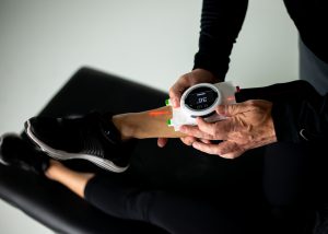
With an absolute new level of assessment and interpretation method, StretchingSA can now offer a full comprehensive program specifically designed for each and every patient’s personal needs.

Our new state of the art technology together with amazing software has turned the tables in physical patient care and quantification of progress.

Enrol with us today, and change your live forever, the right way.

How our musculoskeletal assessments are done:

Most people ask what the assessment entitles before they book their very own first consultation at a StretchingSA branch. Well to enlighten you in this aspect we look at the 5x keys. In our practices we use the skill “clinical” – i.e. competent history taking and examination of our patients – which are the key to making an accurate diagnosis and assessment of you and you’re complaining of joint problems.

The five key questions which needs to be answered are by you:

  • Does the problem arise from the joint, tendon or muscle?
  • Is the condition acute or chronic?
  • Is the condition inflammatory or non-inflammatory?
  • What is the pattern of affected areas/joints?
  • What is the impact of the condition on the patient’s life?

A brief screening examination is done, which takes only 1–2 minutes and has been devised for use in routine clinical assessments. This clinical assessment has been shown to be highly sensitive in detecting significant abnormalities of the musculoskeletal system. It involves inspecting carefully for joint swelling and abnormal posture, as well as assessing the joints for normal movement. A screening examination is known or called as the “GALS’ assessment which normally stands for Gait (overall), Arms, Legs and your Spine.

Fast tell all on assessments:

The Gait assessment:

We normally ask the patient to walk a few steps, turn and walk back, so that we can observe the gait of the patient for symmetry, smoothness and the ability to turn quickly.  The gait assessment is done by asking you/the patient to stain your anatomical position, so that we can assess your shoulder, gluteus, calve muscles, spine and limp alignments.

The Arms assessment:

With the arms assessment we normally ask the patient to place both their hands behind their heads. This gives us an idea of the movement of the shoulder adductions, abductions and forearm rotations. We can also make the assessment of any joint swelling and deformity.

The Legs assessment:

We ask patients to normally lie on our plinths for the legs assessment, by asking you/the patient to do full flexion and extension of both knees. We also assess the internal and external rotation of the hips by helping you guide both the hip and the knee with a 90deg angle for movement. Patients will give us a good indication of joint swelling, deformities as well as callosities of the sole. Most patients will also be assessed for patella effusions and metatarsophalangeal (MTP) joint for inflammatory.


The Spine assessment:

Patients is normally being ask to stand in an upright position so that we can assess for any spine abnormality like scoliosis from side to side and also to check for any other abnormalities like lordosis and kyphosis. We can assess the lateral flexion of the neck from ear to shoulder. Asking to touch your toes, this movement is important to determine the functioning but also to rely on good hip flexion, so it is important for us to palpate for normal movement of the vertebrae.

It is important for us to record both positive and negative findings in the notes of your body. The presence or absence of changes – in appearance or movement – in the gait, arms, legs or spine. With proper musculoskeletal assessment we can always detect if there are abnormalities, swelling of joint etc in your body. This helps us to determine your Stretching protocol and the best way to help you better your living and wellbeing.

So now that you know the basic of the assessment protocol of your first consultation, come and have a pop in or better yet, make your first appointment. You will not be disappointed.


Active isolated stretching

Holding a stretch for 30 seconds and more is BAD !

A study done in Canada, once again showed us that holding your stretches for long periods (45 seconds) is not good for your athletic performance, and even impair your warm up.  If you like to exercise, please don’t warm up with long stretches.

The purpose of the study was to investigate the effect of an acute bout of lower limb static stretching on balance, proprioception, reaction, and movement time.

The conclusion was that an acute bout of static stretching impaired the warm-up effect achieved under control conditions with balance and reaction/movement time.

Methods: Sixteen subjects were tested before and after both a static stretching of the quadriceps, hamstrings, and plantar flexors or a similar duration

control condition. The stretching protocol involved a 5-min cycle warm-up followed by three stretches to the point of discomfort of 45 seconds each with 15-s rest periods for each muscle group. Measurements included maximal voluntary isometric contraction (MVC) force of the leg extensors, static balance using a computerized wobble board, reaction and movement time of the dominant lower limb, and the ability to match 30% and 50% MVC forces with and without visual feedback.

Results: There were no significant differences in the decrease in MVC between the stretch and control conditions or in the ability to match submaximal forces. However, there was a significant (P  0.009) decrease in balance scores with the stretch (2 9.2%) compared with the control (1 17.3%) condition. Similarly, decreases in reaction (5.8%) and movement (5.7%) time with the control condition differed significantly (P  0.01) from the stretch-induced increases of 4.0% and 1.9%, respectively.

Conclusion: In conclusion, it appears that an acute bout of static stretching impaired the warm-up effect achieved under control conditions with balance and reaction/movement time.


Effect of Acute Static Stretching on Force,
Balance, Reaction Time, and Movement Time
DAVID G. BEHM, ANDREW BAMBURY, FARRELL CAHILL, and KEVIN POWER
School of Human Kinetics and Recreation, Memorial University of Newfoundland, St. John’s, Newfoundland, CANADA
ABSTRACT
BEHM, D. G., A. BAMBURY, F. CAHILL, and K. POWER. Effect of Acute Static Stretching on Force, Balance, Reaction Time, and
Movement Time. Med. Sci. Sports Exerc., Vol. 36, No. 8, pp. 1397–1402, 2004. Purpose: The purpose of the study was to investigate
the effect of an acute bout of lower limb static stretching on balance, proprioception, reaction, and movement time. Methods: Sixteen
subjects were tested before and after both a static stretching of the quadriceps, hamstrings, and plantar flexors or a similar duration
control condition. The stretching protocol involved a 5-min cycle warm-up followed by three stretches to the point of discomfort of
45 s each with 15-s rest periods for each muscle group. Measurements included maximal voluntary isometric contraction (MVC) force
of the leg extensors, static balance using a computerized wobble board, reaction and movement time of the dominant lower limb, and
the ability to match 30% and 50% MVC forces with and without visual feedback. Results: There were no significant differences in
the decrease in MVC between the stretch and control conditions or in the ability to match submaximal forces. However, there was a
significant (P  0.009) decrease in balance scores with the stretch (2 9.2%) compared with the control (1 17.3%) condition.
Similarly, decreases in reaction (5.8%) and movement (5.7%) time with the control condition differed significantly (P  0.01) from
the stretch-induced increases of 4.0% and 1.9%, respectively. Conclusion: In conclusion, it appears that an acute bout of stretching
impaired the warm-up effect achieved under control conditions with balance and reaction/movement time. Key Words: STABILITY,
ISOMETRIC FORCE, PROPRIOCEPTION, FLEXIBILITY
Stretching is commonly utilized to increase the range
of motion (ROM) around the joint (12,19) and theorized
to improve athletic performance (28). With the
exception of an increased ROM, recent studies have not
found substantial evidence to support the use of stretching
for improved performance. A number of studies report that
acute and prolonged stretching may actually reduce human
performance through decreases in force (4,14,15) and power
(11,40).
The stretch-induced decreases in force and power have
been attributed to impairments in neural output (2,4,13) as
well as changes to the musculo-tendinous unit (MTU)
(13,34). Fowles et al. (13) demonstrated an increase in
fascicle length of the soleus and lateral gastrocnemius of a
single subject with 30 min of stretching. Studies have reported
both decreases (24,35) and no change (23) in MTU
passive resistance or stiffness with stretching. MTU stiffness
incorporates the muscle, tendon, and other associated
connective tissue and can determine the effectiveness and
rapidity by which internal forces generated by the muscle
are transmitted to the skeletal system (38). Among the
functions of the intrafusal (includes stretch receptors) muscle
fibers, Golgi tendon organs and other proprioceptors is
to aid in the maintenance of balance (26) and detection of
the position of the body in space (proprioception) (8,10).
Acute changes in MTU length, stiffness, force output, and
muscle activation could alter the ability to detect (afferent
proprioception) and respond (efferent muscle activation) to
changes in the immediate environment. Stretch-induced impairments
might affect overall balance and stability or limb
proprioception. Furthermore, a more compliant MTU
(greater muscle and connective tissue slack) in conjunction
with a disturbed activation of the muscle could alter reaction
(RT) and movement (MT) times. There have been no studies
reporting on the effects of an acute bout of stretching on
balance, proprioception or reaction/movement time.
Balance involves the interaction of automatic postural
and voluntary motor commands of both the trunk and limb
musculature (6,30). Automatic postural responses are modulated
by both trunk and leg inputs (5), with the central
nervous system (CNS) performing anticipatory postural adjustments
when expecting self-inflicted postural perturbations
(1). Because under conditions of high instability the
CNS may suppress anticipatory postural adjustments, voluntary
responses of trunk and limb muscles to postural
challenges would play a prominent role. Stretch-induced
changes to either the afferent limb muscle responses (proprioception)
or the mechanical output would be expected to
affect the ability to adapt effectively to stability challenges.
At the elite sport level, where milliseconds can mean the
difference between winning and losing, even small changes
in RT, MT, and balance can have a dramatic impact. For
example, differences between the personal best times of the
Address for correspondence: David Behm, Ph.D., School of Human Kinetics
and Recreation, Memorial University of Newfoundland, St. John’s,
Newfoundland, Canada, A1C 5S7; E-mail: dbehm@mun.ca.
Submitted for publication December 2003.
Accepted for publication April 2004.
0195-9131/04/3608-1397
MEDICINE & SCIENCE IN SPORTS & EXERCISE®
Copyright © 2004 by the American College of Sports Medicine
DOI: 10.1249/01.MSS.0000135788.23012.5F
1397
top sprinters in the world can differ by approximately 1%
(i.e., Greene: 9.79 s, Bailey 9.84 s, Christie: 9.87 s, Cason
9.92 s). Thus, even minor changes in RT, MT, and balance
could have important implications for athletic endeavors.
The possibility of stretch-induced impairments to balance,
RT, and MT not only affects sport applications but the loss
of dynamic balance is also a risk factor for osteoporotic
fractures (27). The contributions of RT and MT to dynamic
balance could have implications not only for athletes and
fitness enthusiasts but also for rehabilitation professionals
who prescribe stretching.
The objective of the present study was to examine alterations
in static balance, proprioception, RT and MT, and
force. It was hypothesized based on previous studies that
demonstrated decreases in force and activation as well as
changes in MTU stiffness that all the dependent variables
would be adversely affected by an acute bout of static
stretching.
METHODOLOGY
Approach to the problem and experimental design.
Because a number of the previous studies investigating
stretch-induced force and power decrements used prolonged
stretching routines (15–30 min) of single muscle
groups (4,13) that were not representative of typical stretching
routines, the present study used a moderate volume of
stretching with three lower-limb muscle groups. Subjects
were tested before and after both an acute bout of static
stretching of the quadriceps, hamstrings, and plantar flexors
or a similar duration control condition. The stretching protocol
involved a 5-min cycle warm-up followed by three
stretches to the point of discomfort of 45 s each with 15-s
rest periods for each muscle group (independent variable).
Measurements were conducted over a 20-min period that
included maximal voluntary isometric contraction (MVC)
force of the leg extensors, static balance using a computerized
wobble board, reaction and movement time of the
dominant lower limb, and the ability to match 30% and 50%
MVC forces with and without visual feedback (dependent
variables).
Subjects. Sixteen healthy male university students (age
 24.1  7.4 yr, weight  71.5  15.4 kg, height  172.3
 6.5 cm) volunteered for the experiment. All participants
were verbally informed of the protocol, and read and signed
a consent form. Each participant also read and signed a
Physical Activity Participation Questionnaire (PAR-Q: Canadian
Society for Exercise Physiology) to ensure that their
health status was adequate for participation in the study. The
study was sanctioned by the Memorial University of Newfoundland
Human Investigations Committee.
Intervention. Before stretching of both legs, subjects
performed a warm-up procedure consisting of a 5-min cycle
on a cycle ergometer (Monark Ergomedic 828E) at 70 rpm
with 1-kp resistance. The order of quadriceps, hamstrings,
and plantar flexors stretching was randomized. Stretches
were held to the threshold of discomfort for a duration of
45 s with 15-s recovery periods between stretches. Each
type of stretch was repeated three times. Stretching of both
legs included a series of unilateral kneeling knee flexion
(quadriceps), hip flexion with extended leg while supine
(hamstrings), extended leg dorsiflexion while standing
(stretch of the plantar flexors with gastrocnemius emphasis),
and flexed knee dorsiflexion while standing (stretch of the
plantar flexors with soleus emphasis). Stretching was passive
for the quadriceps and hamstrings with the same researcher
controlling the change in the range of motion and
resistance for all subjects. The researcher would extend the
limb to the limits of the participant’s range of motion
without incurring injury. Subjects provided their own resistance
for the plantar flexors stretches with the instructions to
stretch the muscles to the point of discomfort.
For the control condition, subjects performed the 5-min
cycle warm-up and were allowed to rest for approximately
26 min between the pre- and posttesting periods. The 26-
min rest period provided similar pre- to posttest durations
for the stretching and control conditions. The order of control
and experimental stretch conditions was randomized.
Testing. An orientation session involving multiple attempts
(minimum three attempts) for all measures was organized
for all subjects 3–5 d before testing. The order of
testing was randomized. The stretching intervention commenced
2 min after the pretesting session. Postintervention
testing began within 1 min after the stretching routine. The
duration of pre- and posttesting was approximately 20 min
each.
For leg extension MVC force, subjects sat on a bench
with hips and knees flexed at 90°, and the upper leg and hips
restrained by two straps. The ankle was inserted into a
padded strap attached by a high-tension wire to a Wheatstone
bridge configuration strain gauge (Omega Engineering
Inc., LCCA 250). Prestretching, subjects performed two
leg extension MVC. If there was more than a 5% difference
in maximum force output, another MVC was performed.
Only two contractions were permitted poststretching to reduce
the chance of fatigue. Three-minute rest periods were
allocated between contractions. The day-to-day reliability of
the strength test using an intraclass correlation coefficient
(ICC) was determined to be 0.9, with a between test (single
session) reliability of 0.93.
All torques were detected by the strain gauge, amplified
(Biopac Systems Inc., DA 100, and analog to digital converter
MP100WSW) and monitored on computer (Sona
Phoenix PC). All data were stored on a computer at a
sampling rate of 2000 Hz. Data were recorded and analyzed
with a commercially designed software program (Acq-
Knowledge III, Biopac Systems Inc.).
The matching force task used the same set-up as the MVC
test. Once the MVC force was established, grid lines were
provided on the computer, which outlined 30% and 50% of
the MVC force. Subjects were asked to exert sufficient
isometric leg extension force over a 5-s period to match the
gridlines. Visual feedback was always given for the first
three trials of a particular relative force (30% or 50% MVC),
while the computer screen was obstructed from view for the
subsequent three trials. Two-minute rest periods were per-
1398 Official Journal of the American College of Sports Medicine http://www.acsm-msse.org
mitted between attempts. The order of the relative force
matching tasks was randomized. The day-to-day reliability
of the matching force test using an ICC was determined
to be 0.8, with a between test (single session) reliability
of 0.88.
A balance ratio (contact with floor to no contact time) was
calculated by a software program (Innervations, Muncie,
IN) from a 30-s wobble board test (Kinematic Measurement
Systems, Muncie, IN). A metal plate connected to the computer
hardware was placed under the wobble board. When
the perimeter of the wobble board made contact with the
metal plate, the duration and frequency (during the 30-s test)
of contact was recorded by the software. Subjects received
an orientation session for the balance board on a separate
day as well as one to two practice attempts on the day of
testing. The day-to-day reliability of the balance test using
an ICC was determined to be 0.81, with a between test
(single session) reliability of 0.86.
RT and MT were measured by an apparatus developed by
the Memorial University of Newfoundland Technical Services
(Electronics, Newfoundland, Canada). The testing apparatus
consisted of a stop clock (58007, Lafayette Instrument
Company, Lafayette, IN), an analog timer (L15–365/
099, Triton Electronics, UK), a stop clock latch (58027,
Lafayette Instrument Company) that connected the stop
clock and the analog timer, a custom-designed box (62 cm
(length)  15.5 cm (width)  9 cm (height)) with the
distance of 50 cm from center of start button to the center of
the stop button, and a trigger plate for the start of the task.
With the device situated on the floor, the task entailed
movement of the dominant foot in response to the illumination
of an incandescent light bulb. The subject would start
with the nondominant foot on the floor and the dominant
foot (ball of foot) placed on the start button. Upon illumination
of the light signal, the subject would release the start
button and move their foot forward to touch the stop button
(50 cm). RT was measured as the time between the illumination
of light stimulus and release of the start button. MT
was measured as the time between the initiation of movement
and the depressing of the stop button. The actions
involved hip flexion, knee extension and plantar flexion. In
order to move as quickly as possible, the quadriceps and
plantar flexors would initiate the movement, while the hamstrings
would aid with the deceleration of the leg to accurately
touch the stop button. Three trials of RT and MT were
performed with 30-s rest periods. The day-to-day reliability
of the RT and MT tests using an ICC was determined to be
0.60 and 0.89 respectively with no significant (P  0.05)
difference between values for test versus retest. Between
test (single session) ICC reliability measures of 0.79 and
0.93 were recorded for RT and MT, respectively.
Statistical analysis. Data were analyzed using a twoway
ANOVA repeated measures design. The factors included
condition (stretching vs control) and testing (pre- and
postcondition). An alpha level of P  0.05 was considered
statistically significant. If significant differences (P  0.05)
were detected, a Bonferroni-Dunn’s procedure was used to
identify the significant change. The means and SEM are
illustrated in Table 1. Reliability was assessed using an
alpha (Cronbach) model ICC (25) with all 16 subjects.
Repeated tests were conducted within 48–72 h.
RESULTS
Overall, significant differences from the control condition
due to the stretching protocol occurred with measures of
static balance, RT and MT.
Force. There was no significant difference between
stretching and control conditions in force output. The stretch
and control conditions experienced similar 6.9% and 5.6%
force decrements, respectively, from the pretest to the
posttest.
Perceived force. Whether visual feedback was or was
not provided, there were no significant differences in the
ability to match 30% and 50% MVC between control and
stretch conditions during the pretest or posttests. The control
condition demonstrated a nonsignificant 18.8% and 10.7%
greater accuracy for maintaining 30% and 50% MVC
posttest.
Balance. Balance scores moved in opposing directions
resulting in a significant change (P  0.009) for the pre- to
posttest differences between control and stretch conditions
(effect size  0.11: small). In comparison with the precontrol
sessions, the control condition demonstrated a significant
(P  0.05) 17.3% improvement in balance scores
postcontrol, whereas the stretch condition showed a nonsignificant
2.2% decrease in balance scores poststretching routine
(Table 1).
Reaction and movement time. Similar to balance
scores, reverse trends for the stretch and control conditions
resulted in significant change for the pre- to posttest differences
with both reaction (P  0.01, effect size  1.11:
TABLE 1. Balance, reaction and movement time data (means  SEM).
Pretest Control
Condition
Posttest Control
Condition
Pre- to Posttest
Difference
Pretest Stretch
Condition
Posttest Stretch
Condition
Pre- to Posttest
Difference
Wobble board contacts 10.8 8.9 1.9 8.8 9.0 0.2
(2.0) (1.5) 917.3%* (1.7) (1.8) 82.2%
Reaction time (RT) 294 ms 277 ms 17 ms 283 ms 294 ms 11 ms
(27.5) (10.7) 95.8% (16.6) (15.8) 84.0%
P  0.16
Movement time (MT) 427 ms 403 ms 24 ms 418 ms 426 ms 8 ms
(37.5) (30.2) 95.7% (32.6) (39.1) 81.9%
P  0.18
* Asterisk indicates a significant difference from the pre-test condition. Significant differences were detected in the pre- to posttest differences between control and stretch conditions
for balance (power: 50%), RT (power: 95%), and MT (power: 50%).
STRETCHING EFFECTS ON BALANCE AND MOVEMENT Medicine & Science in Sports & Exercise 1399
moderate-large) and movement (P  0.01, effect size 
0.65: moderate) time, respectively (Table 1). In reference to
the pretest control session, RT and MT improved (decreased)
by 5.8% (P  0.16) and 5.7% (P  0.18), respectively.
However, compared to the pretest stretch condition,
RT and MT were impaired (increased) by 4.0% and 1.9%
poststretch, respectively (nonsignificant).
DISCUSSION
The most important findings in this study were the impairments
in balance, RT and MT, due to prior stretching.
The control condition which involved a 5-min cycle warmup,
submaximal and maximal leg extension contractions,
three trials each of rapid leg movement (RT and MT), and
balance on a wobble board followed by a 26 min rest period
improved performance in the balance, RT and MT tests.
Inserting a stretching routine within the rest period not only
nullified the beneficial effects of the warm-up but also
produced small performance decrements in relation to the
pretest scores.
These decrements reflect impairments associated with
recent studies that have reported stretch-induced decreases
in force (4,13,14), power (11,40), and muscle activation
(2,4,13). Although isometric forces decreased 6.9% after
stretching in the present study, the decrement was not significantly
greater than the 5.6% impairment in the control
condition. The lack of a significant loss of isometric force
may be attributed to the moderate volume of stretching
imposed. In contrast to other similar studies that have
stretched a single muscle group for 15–30 min (4,13,14), the
present study involved only 135 s of intermittent stretching
for each of the three muscle groups.
Balance involves the interaction of automatic postural
and voluntary motor commands of both the trunk and limb
musculature (6,30). Balancing on a wobble board can involve
unanticipated perturbations to equilibrium that are
adjusted through contractions of both trunk and limb muscles.
Bloem and colleagues (6) speculated that lower leg
inputs act to modulate automatic postural responses. They
also found that the knees, hips, and trunk initiated movement
before the automatic postural responses. The CNS
performs anticipatory postural adjustments when expecting
self-inflicted postural perturbations (1). However, Aruin and
colleagues (1) suggested that under conditions of high instability
that the CNS may suppress anticipatory postural
adjustments as protection against their possible destabilizing
effects. Consequently, voluntary responses of trunk and
limb muscles to postural challenges would play a prominent
role. Shiratori and Latash (30) in a subsequent study from
the same laboratory reported that distal muscles (tibialis
anterior and soleus) cope with asymmetrical perturbations
and modulate the anticipatory postural adjustments in novel
situations (i.e., wobble board). Furthermore, Lipshits et al.
(22) described how perturbing balance by rapidly raising a
hand was initially counteracted by activation of lower limb
muscles. Therefore, it is apparent the important role that
lower limbs play in maintaining balance. Modifications to
either the afferent limb muscle responses or the mechanical
output would be expected to affect the ability of the peripheral
neuromuscular system to adapt effectively to stability
challenges.
Stretching has been reported to alter the length and stiffness
of the affected limb MTU. Although the exact mechanisms
responsible for increases in range of motion after
stretching are debatable, the increase is commonly attributed
to decreased MTU stiffness (37,39). Fowles et al. (13)
demonstrated an 8-mm increase in fascicle length of the
soleus and lateral gastrocnemius with 30 min of stretching.
Studies have reported both decreases (24,35) and no change
(23) in MTU passive resistance or stiffness with stretching.
Changes in MTU stiffness might be expected to affect the
transmission of forces, the rate of force transmission and the
rate at which changes in muscle length or tension are detected.
A more slack parallel and series elastic component
could increase the electromechanical delay by slowing the
period between myofilament crossbridge kinetics and the
exertion of tension by the MTU on the skeletal system. In
addition, the detection and monitoring of the muscle tension
by the Golgi tendon organs (GTO) would be delayed since
a more compliant tendon would not transmit the tension
information to the GTO as rapidly as a stiffer MTU. Furthermore,
increases in MTU length and decreases in MTU
stiffness could also alter the perception of the intrafusal
stretch receptors and thus perturb the afferent responses to
both changes in muscle length, rate of length change, and
tension (GTO). Therefore, stretch-induced changes in muscle
compliance might affect both the muscle afferent input
to the CNS and muscle output for counteracting unexpected
perturbations to balance.
Further evidence for the detrimental effect of an acute
bout of stretching on the CNS has been provided by Avela
et al. (2). They investigated the effects of passive stretching
of the triceps surae muscle on reflex sensitivity. After 1 h of
stretching, there were significant decreases in MVC
(23.2%), EMG (19.9%), stretch reflex peak-to-peak amplitude
(84.8%), and the ratio of H-reflex to muscle compound
action potential (M-wave) (43.8%). Although neural propagation
seemed unaffected (M-wave), afferent excitation of
the motoneuron pool (H-reflex) was impaired. They suggested
that the decrease in the excitation of the motoneuron
pool resulted from a reduction in excitatory drive from the
Ia afferents onto the -motoneurons, possibly due to decreased
resting discharge of the muscle spindles via increased
compliance of the MTU.
Stretch-induced impairments in RT and MT may be related
to similar mechanisms as the disturbance in balance.
As mentioned previously, a more compliant MTU could
compromise the rate of tension development. Although it is
highly unlikely that the visual detection of the light stimulus
and the subsequent initiation of CNS motor programs to
move the leg would be adversely affected by stretching, a
prolonged electromechanical delay could negatively affect
both RT and MT. Although not monitored in the present
study, other studies have reported decreases in muscle activation
after stretching (2,4,13). Increases in motoneuron
1400 Official Journal of the American College of Sports Medicine http://www.acsm-msse.org
inhibition are more likely to affect the high-threshold fastcontracting
motor units that could also play a role in stretchinduced
RT and MT impairments.
An interesting development in the present study was the
control condition’s improvements in balance scores, RT and
MT. This finding may provide support for the beneficial
effect of a short duration, combination of general (cycle
warm-up and leg extension contractions) and specific (pretest
wobble board, RT and MT tests) activities. However,
because there was no condition in which a cycle warm-up
was not included, the contribution of the cycling cannot be
precisely deduced from the present study.
Young and Behm (41) reported similar results in a study
where subjects participated in five different warm-ups in a
randomized order before the performance of two jumping
tests. The warm-ups were: a) control, b) 4-min run, c) static
stretch, d) run and static stretch, and e) run and static stretch
and practice jumps. Generally, the stretching warm-up produced
the lowest values and the run or run and stretch and
jumps warm-ups produced the highest values of explosive
force production. Thus, it should not be surprising that the
control condition’s dynamic warm-up and static leg extension
contractions facilitated subsequent performance.
Numerous studies have investigated the effects of actively
warming-up on subsequent performance, yielding
mixed results. Although the majority of the research has
demonstrated that an increase in temperature facilitates human
performance (9,29,32), other studies have shown inhibitory
effects (5,16) as well as no effect (7) of warming-up
on subsequent performance. These conflicting results may
be attributed to discrepancies in the type of exercise, intensity,
duration, or any combination of these variables utilized
in the warm-up procedure. Studies have demonstrated that
warming-up can result in increased nerve conduction velocity
(31). Increases in nerve conduction velocity could facilitate
the response speed to perturbation in balance as well as
contributing to the improvements in RT and MT.
Another mechanism that may help explain the control
condition’s improvement in RT, MT, and balance may be
the effect of postactivation potentiation (PAP). PAP can be
defined as an increase in the efficiency of the muscle to
produce submaximal force after a voluntary contraction.
PAP has been attributed to regulatory light chain (RLC)
phosphorylation (17,20,21,33), which increases the number
of force-producing crossbridges under conditions of suboptimal
Ca2 activation (33). Suboptimal Ca2 activation may
be present with lower-frequency stimulation such as the
lower-intensity contractions associated with static balance.
Potentiation also involves an increase in the rate constant of
crossbridge attachment (20). The increased rate constant
would allow a greater number of crossbridges to form during
a specific time period resulting in increased force and
rate of force development capabilities. Furthermore, at the
supraspinal level, motor-evoked potential facilitation has
been reported after different durations (5, 15, and 30 s) and
intensities (10%, 25%, and 50%) of thenar muscle voluntary
contractions (3). A number of studies have suggested that an
improved neuromuscular activation can occur after a few
MVC (18,40). Evidence of this postcontraction neural potentiation
is provided by increased H-reflex amplitudes (18)
that may persist for 10 min after the contractions (36). Thus,
pretest contractions in the control condition may have elicited
a PAP response providing both a facilitation of the
motoneuron excitation and RLC phosphorylation contributing
to the significant improvements in RT and MT. Indirectly,
the PAP-induced augmentation of RT and MT would
also benefit balance by allowing more rapid responses to the
perturbations of the unstable environment. The stretching
condition may have nullified the beneficial effects of PAP
contributing to the 2.2% decrement in balance scores.
ICC (reliability) for the dependent measures were all in
the good to excellent category (0.80–0.93) except for the
day-to-day reliability of RT that scored 0.60 (moderate). A
paired samples t-test was then conducted on the RT measures.
The lack of significant difference between the measures
suggested that the low RT ICC may be attributed to the
low between subject variability. Another contributing factor
for this less than optimal reliability may be due to the test
set-up. The RT test for the lower limb necessitated that the
individuals place most of their mass on the nondominant
limb creating a degree of instability. Even with an orientation
session, the lack of familiarity with this type of movement
and the greater instability may have led to a less
consistent action.
CONCLUSION
In summary, the findings of the present study demonstrate
that a moderate bout of stretching (three repetitions per
muscle group) held to the point of discomfort can adversely
affect performance on tests of static balance, RT and MT.
The stretch-induced impairments are hypothesized to be
related to changes in muscle compliance with the stretching
that may adversely affect the ability to detect and respond to
changes in muscle length, and rate of change in muscle
length and forces. Furthermore, it was found in the present
study that a warm-up consisting of general and specific
activities related to the tasks may improve performance
even after 20 min of recovery. Considering the minute
differences between winning and losing in both individual
and team sports as well as the precarious balance or stability
of the elderly, the low but significant percentage changes in
RT, MT, and balance could result in serious consequences.
REFERENCES
1. ARUIN, A. S., W. R. FORREST, and M. L. LATASH. Anticipatory
postural adjustments in conditions of postural instability. Electroencephal.
Clin. Neurophysiol. 109:350–359, 1998.
2. AVELA, J., H. KYR¨OL¨AINEN, and P. V. KOMI. Altered reflex sensitivity
after repeated and prolonged passive muscle stretching.
J. Appl. Physiol. 86:1283–1291, 1999.
STRETCHING EFFECTS ON BALANCE AND MOVEMENT Medicine & Science in Sports & Exercise 1401
3. BALBI, P., A. PERRETTI, M. SANNINO, L. MARCANTONIO, and L.
SANTORO. Post-exercise facilitation of motor evoked potentials
following transcranial magnetic stimulation: a study in normal
subjects. Muscle Nerve 25:448–452, 2002.
4. BEHM, D. G., D. BUTTON, and J. BUTT. Factors affecting force loss
with stretching. Can. J. Appl. Physiol. 26:262–272, 2001.
5. BISHOP, D., D. BONETTI, and B. DAWSON. The effect of three
different warm-up intensities on kayak ergometer performance.
Med. Sci. Sports Exerc. 33:1026–1032, 2001.
6. BLOEM, B. R., J. H. J. ALLUM, M. G. CARPENTER, and F. HONEGGER.
Is lower leg proprioception essential for triggering human automatic
postural responses? Exp. Brain Res. 130:375–391, 2000.
7. BRUYN-PREVOST, P. The effect of various warming-up intensities
and durations upon some physiological variables during an exercise
corresponding to PWC 170. Eur. J. Appl. Physiol. 43:93–100,
1980.
8. BURKE, D. Muscle spindle function during movement. In: The
Motor System in Neurobiology, E. V. Evarts, S. P. Wise, and D.
Bousfield (Eds.). New York: Elsevier Biomedical Press, 1985, pp.
168–172.
9. CHWALBINSKA-MONETA, J., and O. HANNINEN. Effects of active
warming-up on thermoregulatory, circulatory, and metabolic responses
to incremental exercise in endurance trained athletes. Int.
J. Sport Med. 10:25–29, 1989.
10. COOKE, J. D. The role of stretch reflexes during active movements.
Brain Res. 181:493–497, 1980.
11. CORNWELL, A., A. G. NELSON, and B. SIDAWAY. Acute effects of
stretching on the neuromechanical properties of the triceps surae
muscle complex. Eur. J. Appl. Physiol. 86:428–434, 2002.
12. FERBER, R., L. R. OSTERNIG, and D. GRAVELLE. Effect of PNF
stretch techniques on knee flexor muscle EMG activity in older
adults. J. Electromyogr. Kinesiol. 12:391–397, 2002.
13. FOWLES, J. R., D. SALE, and J. D. MACDOUGALL. Reduced strength
after passive stretch of human plantar flexors. J. Appl. Physiol.
89:1179–1188, 2000.
14. FOWLES, J. R., and D. G. SALE. Time course of strength deficit after
maximal passive stretch in humans. Med. Sci. Sports Exerc. 29:
S26, 1997.
15. FOWLES, J. R., D. G. SALE, and J. D. MACDOUGALL. Reduced
strength after passive stretch of the human plantar flexors. J. Appl.
Physiol. 89:1179–1188, 2000.
16. GENOVELY, H., and B. A. STAMFORD. Effects of prolonged warm-up
exercise above and below the anaerobic threshold on maximal
performance. Eur. J. Appl. Physiol. 48:232–330, 1982.
17. GRANGE, R. W., R. VANDENBOOM, and M. E. HOUSTON. Physiological
significance of myosin phosphorylation in skeletal muscle.
Can J. Appl. Physiol. 18:229–242, 1993.
18. GULLICH, A., and D. SCHMIDTBLEICHER. MVC-induced short-term
potentiation of explosive force. New Studies in Athletics 11:67–
81, 1996.
19. HARVEY, L., R. HERBERT, and J. CROSBIE. Does stretching induce
lasting increases in ROM? A systematic review. Physiotherapy
Res. Intern. 7:1–13, 2002.
20. HOUSTON, M. E., and R. W. GRANGE. Myosin phosphorylation,
twitch potentiation, and fatigue in human skeletal muscle. Can
J. Physiol. Pharm. 68:908–913, 1990.
21. HOUSTON, M. E., H. J. GREEN, and J. T. STULL. Myosin light chain
phosphorylation and isometric twitch potentiation in intact human
muscle. Pflu¨gers Arch. Eur. J. Physiol. 403:348–352, 1985.
22. LIPSHITS,M. I., K.MAURITZ, and K. E. POPOV. Quantitative analysis
of anticipatory postural components of a complex voluntary movement.
Fiziologiya Cheloveka 7:411–419, 1982.
23. MAGNUSSON, S. P., P. AAGAARD, and J. J. NIELSEN. Passive energy
return after repeated stretches of the hamstring muscle tendon unit.
Med. Sci. Sports Exerc. 32:1160–1164, 2000.
24. MAGNUSSON, S. P., E. B. SIMONSEN, P. AAGAARD, and M. KJAER.
Biomechanical responses to repeated stretches in human hamstring
muscle in vivo. Am. J. Sports Med. 24:622–627, 1996.
25. MCGRAW, K. O., and S. P. WONG. Forming inferences about some
intraclass correlation coefficients. Psychol. Methods 1:30–46,
1996.
26. NASHNER, L. M. Adapting reflexes controlling the human posture.
Exp. Brain Res. 26:59–72, 1976.
27. NELSON, M. E., M. A. FIATORONE, C. M. MORGANTI, I. TRICE, R. A.
GREENBERG, and W. J. EVANS. Effects of high intensity strength
training on multiple risk factors for osteoporotic fractures: a randomized
control trial. JAMA 272:1909–1914, 1994.
28. POLIDORO, J. R. Sport and Physical Activity in the Modern World.
Needham Heights, MA: Allyn and Bacon, 2000, pp. 124–132.
29. ROBERGS, R. A., D. D. PASCOE, D. L. COSTILL, et al. Effects of
warm-up on muscle glycogenolysis during intense exercise. Med.
Sci. Sports Exerc. 23:37–43, 1991.
30. SHIRATORI, T., and M. LATASH. The roles of proximal and distal
muscles in anticipatory postural adjustments under asymmetrical
perturbations and during standing on rollerskates. Clin. Neurophysiol.
111:613–623, 2000.
31. STEGMAN, D. F., and J. P. DE WEERD. Modelling compound action
potentials of peripheral nerves in situ. II. A study of the influence
of temperature. Electroencephal. Clin. Neurophysiol. 54:516–529,
1982.
32. STEWART, I. B., and G. G. SLIEVERT. The effect of warm-up intensity
on range of motion and anaerobic performance. J. Orthop.
Sports Phys. Therapy 27:154–161, 1998.
33. SWEENEY, H. L., B. F. BOWMAN, and J. T. STULL. Myosin light chain
phosphorylation in vertebrate striated muscle: regulation and function.
Am. J. Physiol. (Cell Physiol.) 264:c1085–c1095, 1993.
34. TAYLOR, D. C., J. D. DALTON, A. V. SEABER, and W. E. GARRET.
Viscoelastic properties of muscle-tendon units: the biomechanical
effects of stretching. Am. J. Sports Med. 18:300–308, 1990.
35. TOFT, E., G. T. ESPERSEN, S. KåLUND, T. SINKJÆR, and B. C.
HORNEMANN. Passive tension of the ankle before and after stretching.
Am. J. Sports Med. 17:489–494, 1989.
36. TRIMBLE, M. H., and S. S. HARP. Postexercise potentiation of the
H-reflex in humans. Med. Sci. Sports Exerc. 30:933–941, 1998.
37. WILSON, G., B. ELLIOT, and G. WOOD. Stretching shorten cycle
performance enhancement through flexibility training. Med. Sci.
Sports Exerc. 24:116–123, 1992.
38. WILSON, G. J., A. MURPHY, and J. F. PRYOR. Musculotendinous
stiffness: its relationship to eccentric, isometric and concentric
performance. J. Appl. Physiol. 76:2714–2719, 1994.
39. WILSON, G. J., G. A. WOOD, and B. C. ELLIOT. The relationship
between stiffness of the musculature and static flexibility: an
alternative explanation for the occurrence of muscular injury.
Intern. J. Sports Med. 12:403–407, 1991.
40. YOUNG, W., and J. ELLIOTT. Acute effects on static stretching,
proprioceptive neuromuscular facilitation stretching, and maximum
voluntary contractions on explosive force production and
jumping performance. Res. Q. Exerc. Sport 72:273–279, 2001.
41. YOUNG, W. B., and D. G. BEHM. Effects of running, static stretching
and practice jumps on explosive force production and jumping
performance. J. Sport Med. Phys. Fitness 34: 119–124, 2003.
1402